Besides computer vision methods, the newest data science techniques, such as artificial intelligence, are being used at VITO to retrieve information from retinal images. Artificial intelligence is used to determine patterns in images and uses GPU processors for intense calculations.
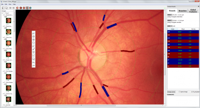
Blood vessels in the retina of the eye show strong similarities with the blood vessels of the central circulatory system and those in the brain. They also react in the same manner to risk factors for cardiovascular diseases. Modifications in the dimensions and structure of the blood vessels in the retina can be used as a biomarker for what occurs in the rest of the body. Image analysis of the retina can therefore be used to assess the risk of developing chronical diseases (hypertension, myocardial infarction, stroke, diabetes and even Alzheimer’s disease) and to monitor the evolution of these diseases. IFLEXIS is a software that enables rapid analyses of the pattern of the blood vessels using digital retinal images.
Window to the heart and the brain
The retina develops from the brain as a layer with light-sensitive cells behind the eye which allows us to see. The circulation of the retina is assured by very small blood vessels. With the help of so-called retinal cameras, digital images of these blood vessels can be made in a non-invasive and simple manner.
More than 2000 years ago Confucius already knew that “A picture says more than a thousand words”. One single image can provide more information in a split second than dozens of pages. The same applies to medical images – like images of the retina. An experienced researcher or doctor interprets these images at a glance and gives a significance to the image. The quality and outcome depends not only on the quality of the image, but also on the experience of the expert.
In practice, retinal images are taken at an ophthalmologist for the detection and assessment of medical conditions of the retina like diabetic retinopathy and glaucoma. However, an in-depth quantitative analysis of the exposed blood vessel structure does not take place. Nevertheless, this can be of relevance to identify the first indications of these conditions. Furthermore, the retinal blood vessel pattern is very similar to the central vascular system and brain. Modifications in the thickness and pattern of those blood vessels are considered as early indicators of the development and progression of chronic diseases. Several extensive epidemiological studies show that deviations in the retinal blood vessels could predict the risk of chronic diseases. There are already indications for hypertension, myocardial infarction and stroke.
IFLEXIS software
Retina analyses are applied in VITO’s healthcare technology research. On the one hand, the application of retina technology is promoted in research, on the other hand there is more technological focus on the development of new algorithms and data science techniques to retrieve more information from digital retinal images.
In this context, IFLEXIS software is developed. The software allows accurate quantification of the blood vessel pattern. Dimensions of oxygen-rich (arterioles) and oxygen-deficient (venules) are being calculated, as well as branching patterns of individual blood vessels and the entire network of blood vessels. The software converts a retinal image into more than 30 parameters. This is ideal to determine modifications in the structure of the blood vessels and to detect potential biomarkers. The first software licenses have already been sold. The promotion of IFLEXIS is underway with later this year scientific and commercial contributions on large international conferences like ARVO and EUretina.
The current software is primarily intended for study and research purposes in ophthalmology, cardiology, neurology and epidemiology. New software developments, focusing on fully automised analysis and extraction of more information from the images, is underway. The first steps towards CE-marking are ongoing. All this should lead to a product that has a multiplicity of uses; which will significantly increase the market potential.
Image analysis with artificial intelligence
Besides computer vision methods, the newest data science techniques, such as artificial intelligence, are being used at VITO to retrieve information from retinal images. Artificial intelligence is used to determine patterns in images and uses GPU processors for intense calculations.
At the moment, an application is being refined for the recognition of the blood vessel pattern. A second application focuses on the screening to the stages of diabetic retinopathy. This clinical complication of diabetes is one of the leading causes of blindness worldwide. An early detection could prevent blindness. A computer model is established based on a training set of tens thousands of labeled images. The first results in identifying the different stages of the disease are promising. Analysis of independent data sets indicates that the model approaches the performance of a specialist doctor. The experience acquired is now also being used to identify other medical problems of the retina. Researchers at home and abroad are working on this topic. During the research, immediate identification of the key economic and social values of such applications takes place.


